
具体描述
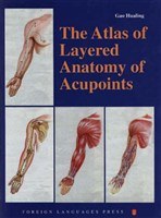
针灸穴位层次解剖图谱,ISBN:9787119017532,作者:高华龄
作者简介
Gao Hualing, bom in 1926, is a
native of Wangkui County,
Heilongjiang Province. He graduated
from the Medical Department of the
Chinese Medical Sciences University
in 1948, and finished his postgraduate
studies at the Beijing Union Medical
College in 1956. After graduation, he
taught at the Changchun No. 3 Military
Medical Sciences University, and the
Changchun No. 1 Military Medical
Sciences University, and later worked
as a lecturer, associate professor,
professor, tutor of graduate students,
and head of the Human Anatomy
Teaching and Research Office of the
Beijing University of Traditional
Chinese Medicine. Gao Hualing is a
member of the Acupuncture Society,
and the Anatomy Society under the
Chinese Medical Association as well as
a member of the China Association of
Senior Professors. He has made the
teaching and study of human anatomy
his lifetime's work, and has published
many papers, including "Observations
of Arteria Subclavia in Chinese
People," "A Study of Arteria Axillaris
in Chinese People," "A Study of
Chinese Women's Breast Arteria," "A
Study of Chinese Women's Womb
Arteria," and "A Study of the Needle
Direction for Acupuncture Analgesia."
目录信息
Preface
Part 1
Fig. 1(1) The nomenclature, gauge and diameter of the needle appar-
atuses
Fig. 1 (2) The cun and finger measurement on body for acupuncture-
moxibustion
Fig. 2(1) The commonly used postures of the patient for acupuncture-
moxibustion
Fig. 2 (2) The directions for needle insertion
Fig. 3 The osseous and muscular landmarks on the anterior surface of
the body
Fig. 4 The osseous and muscular landmarks on the posterior surface of
the body
Fig. 5 The osseous and muscular landmarks on the lateral surface of
the body
Fig. 6 The segmental skin nerve distribution on the anterior aspect of
the body
Fig. 7 The segmental skin nerve distribution on the posterior aspect of
the body
Appendix 1: The courses, the nomenclature of the points and indications
of the fourteen meridians
Part2
Fig. 8 The skin and points on the craniofacial region
Fig. 9 The muscles and points on the craniofacial region
Fig. 10 The blood vessels, nerves and points on the craniofacial region
Fig. 11 The skeleton and points on the craniofacial region
Fig. 12 The skin and points on the posterior of head
Fig. 13 The muscles and points on the posterior of head
Fig. 14 The blood vessels, nerves and points on the posterior of head
Fig. 15 The skeleton and points on the posterior of head
Fig. 16 The skin and points on the right aspect of the cephalocervix
Fig. 17 The muscles and points on the right aspect of the cephalocervix
Fig. 18 The blood vessels, nerves and points on the right aspect of the
cephalocervix
Fig. 19 The skeleton and points on the right aspect of the cephalocervix
Fig. 20 The skin and points on the anterior aspect of the trunk
Fig. 21 The muscles and points on the anterior aspect of the trunk
Fig. 22 The blood vessels, nerves and points on the anterior aspect of
the trunk
Fig. 23 The organs and points on the anterior aspect of the trunk
Fig. 24 The skeleton and points on the anterior aspect of the trunk
Fig. 25 The skin and points on the posterior aspect of the trunk
Fig. 26 The muscles and points on the posterior aspect of the trunk
Fig. 27 The blood vessels, nerves and points on the posterior aspect of
the trunk
Fig. 28 The organs and points of the posteiror aspect of the trunk
Fig. 29 The skeleton and points on the posterior aspect of the trunk
Fig. 30 The skin and points on the right aspect of the trunk
Fig. 31 The muscles and points on the right aspect of the trunk
Fig. 32 The blood vessels, nerves and points on the right aspect of the
trunk
Fig. 33 The organs and points on the right aspect of the trunk
Fig. 34 The skeleton and points on the right aspect of the trunk
Fig. 35 The skin and points on the anterior aspect of the right upper
limb
Fig. 36 The muscles and points on the anterior aspect of the right upper
limb
Fig. 37 The blood vessels nerves and points on the anterior aspect of
the right upper limb
Fig. 38 The skeleton and points on the anterior aspect of the right upper
limb
Fig. 39 The skin and points on the posterior aspect of the right upper
limb
Fig. 40 The muscles and points on the posterior aspect of the right
upper limb
Fig. 41 The blood vessels, nerves and points on the posterior aspect of
the right upper limb
Fig. 42 The skeleton and points on the posterior aspect of the right
upper limb
Fig. 43 The skin and points on the lateral aspect of the right upper limb
Fig. 44 The muscles and points on the lateral aspect of the right upper
limb
Fig. 45 The blood vessels, nerves and points on the lateral aspect of the
right upper limb
Fig. 46 The skeleton and points on the lateral aspect of the right upper
limb
Fig. 47 The skin and points on the anterior aspect of the right lower
limb
Fig. 48 The muscles and points on the anterior aspect of the right lower
limb
Fig. 49 The blood vessels nerves and points on the anterior aspect of
the right lower limb
Fig. 50 The skeleton and points on the anterior aspect of the right lower
limb
Fig. 51 The skin and points on the posterior aspect of the right lower
limb
Fig. 52 The muscles and points on the posterior aspect of the right
lower limb
Fig. 53 The blood vessels nerves and points on the posterior aspect of
the right lower limb
Fig. 54 The skeleton and points on the posterior aspect of the right
lower limb
Fig. 55 The skin and points on the lateral aspect of the right lower limb
Fig. 56 The muscles and points on the lateral aspect of the right lower
limb
Fig. 57 The blood vessels, nerves and points on the lateral aspect of the
right lower limb
Fig. 58 The skeleton and points on the lateral aspect of the right lower
limb
Fig. 59 The skin and points on the medial aspect of the right lower limb
Fig. 60 The muscles and points on the medial aspect of the right lower
limb
Fig. 61 The blood vessels, nerves and points on the medial aspect of the
right lower limb
Fig. 62 The skeleton and points on the medial aspect of the right lower
limb
Fig. 63 (1) The skin and points on the anterior aspect of the auricle
Fig. 63 (2) The veins and points on the anterior aspect of the auricle
Fig. 63 (3) The arteries, nerves and points on the anterior aspect of the
auricle
Fig. 64 (1) The skin and points on the posterior aspect of the auricle
Fig. 64 (2) The arteries nerves and points on the posterior aspect of
the auricle
Fig. 64 (3) The veins and points on the posterior aspect of the auricle
Fig. 65 (1) The distribution of the points on the anterior aspect of
the auricle
Fig 65 (2) The distribution of the points on the posterior aspect of the
auricle
Appendix 2: The points of the fourteen meridians, the location, the
posture of the patient, the direction, angle and depth of insertion
of the needle, and the indications
Appendix 3: Precautions in acupuncture treatment
Appendix 4: The selected points in acupuncture treatment of common
diseases
· · · · · · (收起)
读后感
评分
评分
评分
评分
用户评价
这本书的价值,在我看来,不仅在于它展示了“哪里有穴位”,更在于它阐释了“为什么这个地方有效”。它打破了传统教学中对穴位孤立存在的认知,将所有的点都置于一个动态的、相互关联的生理网络中去考察。我特别喜欢其中关于“特定穴位对自主神经系统调控”的章节。作者没有停留在描述症状的改善,而是深入挖掘了刺激特定穴位后,交感和副交感神经活动的变化趋势,甚至引用了电生理学的研究数据来支撑论点。这种跨学科的整合视角,极大地拓宽了我们对传统针灸理论的理解边界,使其更符合现代医学的认知框架。这对于那些希望将传统疗法融入现代综合治疗方案的临床医生来说,提供了极强的理论支撑和实践信心。
评分读完这本书,我最大的感受是如释重负,因为它解决了我长期以来在教学和临床中遇到的一个核心难题:如何系统化地梳理和传授那些看似庞杂的穴位知识。这本书的逻辑结构安排得极为巧妙,它没有采用简单的按经络顺序罗列,而是根据功能主题进行了模块化划分,这使得知识点的内聚性大大增强。例如,关于“镇静安神”主题的穴位群,它将来自不同经络但作用机制相似的穴位集合在一起进行对比分析,这种对比教学法非常高效。更妙的是,书中还加入了大量的“临床误区辨析”小节,这些内容往往是实践中最容易出错的地方,作者用一种过来人的口吻进行提点,非常贴心,减少了读者走弯路的风险。
评分我必须承认,我最初被这本书吸引,是冲着它“图谱”的名头去的,期望能找到一些高清、直观的解剖插图。然而,这本书给我的惊喜远超预期。它的图文排版设计简直是一流的艺术品,每一个插图都像是精心雕琢的艺术品,线条的粗细、色彩的运用都精准地服务于信息传达的目的。比如,在描述深层刺激技术时,那些三维透视图简直是教科书级别的范例,清晰地展示了针尖在不同深度下可能遇到的肌肉层次、筋膜层以及脏器投影区域。我过去在某些操作上感到犹豫不决的地方,仅通过研读这本书中的几个关键图示和配文,便豁然开朗。这种将精确解剖学知识与临床操作技巧无缝结合的能力,是很多同类书籍望尘莫及的。它成功地将“手上的感觉”和“眼前的影像”在纸面上统一起来,极大地提升了学习和回顾的效率。
评分这本书的装帧和纸张质量也值得称赞,这在医学专业书籍中并不常见。厚实的铜版纸使得那些复杂的彩色图谱能够完美呈现细节,即便是经过多次翻阅和携带,书本的形态依然保持得很好,这对于需要经常带到工作场所作为参考的书籍来说,是至关重要的实用性考量。从设计角度看,页边距的留白恰到好处,既保证了阅读的舒适度,又为读者留出了足够的批注和标记空间。我习惯在书边写下我个人的临床心得或与其他理论的联想,这本书的设计完全支持这种互动式的学习过程,使得它在使用过程中,逐渐被我个人的经验所“个性化”,成为了一本真正属于我自己的工具书,而非仅仅是一本冰冷的教材。
评分这部著作的深度和广度确实令人惊叹,它不仅仅是一本关于经络和穴位的工具书,更像是一部融合了传统智慧与现代科学的桥梁。我花了很长时间研究其中关于特定病症的循证医学证据部分,发现作者在梳理历代医家论述的同时,也非常谨慎地引入了最新的影像学和生物力学研究成果来佐证穴位刺激的实际作用机制。例如,书中对“合”穴的讲解,不仅限于其在经络系统中的功能定位,还深入探讨了这些穴位皮下组织结构(如神经末梢、血管丛)的特殊性,这一点对于临床操作的精准性至关重要。阅读过程中,我尤其欣赏作者在行文风格上保持的那种严谨又不失启发性的平衡感。它不会用过于晦涩的术语堆砌来制造距离感,而是用清晰的逻辑链条引导读者,一步步揭示人体复杂系统中的微妙关联。对于初学者来说,它提供了坚实的基础框架;对于资深从业者,它无疑是一部值得反复研读的案头必备参考,每一次翻阅都能带来新的感悟和操作上的启发。
评分 评分 评分 评分 评分相关图书


本站所有内容均为互联网搜索引擎提供的公开搜索信息,本站不存储任何数据与内容,任何内容与数据均与本站无关,如有需要请联系相关搜索引擎包括但不限于百度,google,bing,sogou 等
© 2026 book.wenda123.org All Rights Reserved. 图书目录大全 版权所有