
具体描述
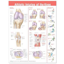
Athletic Injuries of the Knee is designed as a tool to help primary care and sports medicine practitioners and therapists explain anatomical and sports injury concepts to their patients and clients. This chart provides an overview of normal knee anatomy and common injuries and showcases 11 images which illustrate the mechanisms of knee injuries in the context of a human figure playing sports. The vibrant images are from the Anatomical Visual Guide to Sports Injuries and are listed below: Pathological Knee Injury Images: LCL Tear MCL Tear ACL Tear PCL Tear Patellar Tendinopathy: Shows tendinopathy at the following sites: distal quadriceps femoris tendon, distal pole of patella, patellar tendon insertion onto the tibial tubercle Meniscus Tears: Shows bucket handle tear, vertical tear, radial tear, parrot beak tear, fraying/degenerative Sports Injury Mechanism Images: ACL Tear: Basketball ACL Tear: Skiing PCL Tear: Wrestling MCL Tear: Football LCL Tear: Rugby Hyperflexion/Meniscus Tear: Skating Jumper's Knee: Volleyball Patellar Tendon Rupture: Weight Lifting Tibia Fracture: Soccer IT Band Syndrome: Running Normal Anatomy Images: Anterior View of Knee Medial View of Knee Superior view of Knee Showing Meniscus
作者简介
目录信息
读后感
评分
评分
评分
评分
用户评价
这本书的装帧和纸张质量堪称一流,这对于一本需要反复翻阅和比对的工具书来说是至关重要的。我担心如果是一般的光滑纸张,在标记和涂写笔记时可能会打滑,但这里的纸张处理得恰到好处,既保证了图像的色彩还原度,又具备了良好的书写手感。我注意到一个细节,那就是在处理一些深层结构,比如韧带的交叉点时,作者似乎采用了某种特殊的着色技巧,使得结构之间的层次感非常清晰,没有出现“一团糟”的重叠感。这对于理解交叉韧带群的复杂空间关系帮助极大。我过去在其他资料中常常被这些复杂的立体结构搞得晕头转向,但在这里,一切都变得井井有条。此外,书中对常见手术入路或关节镜检查视野的模拟图也相当精彩,这对于正在学习介入性治疗技术的学生来说,提供了宝贵的预习材料。它将静态的解剖学知识与动态的临床操作紧密地结合了起来,体现了作者不仅是解剖学家,更是一位深谙临床实践的专家。
评分如果要用一个词来形容这本书给我的感受,那就是“权威性”。从线条的精确度到术语的使用规范,都透露出一种不容置疑的专业气息。我特别留意了书中所引用的骨性标志和软组织附着点,它们与我熟悉的骨骼学标准完全吻合,这确保了图谱在测量和定位上的高可靠性。这本书的价值远超出一本普通的插图集,它更像是一个跨越了数十年临床观察和解剖研究的浓缩精华。我敢肯定,无论我未来的专业方向是骨科、运动医学还是物理治疗,这本书都会是我书架上最常被取下的那一本。它没有故作高深地使用过于花哨的排版技巧,而是选择了最朴素、最有效的方式——用无可挑剔的解剖图说话。对于任何一个将膝关节解剖视为职业基石的人来说,投资这本书绝对是一笔物超所值的投入,它提供的知识深度和广度,在同类出版物中几乎找不到对手。
评分说实话,我原本对手册类的图集总抱着一种审慎的态度,总觉得它们要么过于简化,要么就是充满了晦涩难懂的术语。然而,这本关于膝盖损伤的图册彻底颠覆了我的看法。它成功地在学术严谨性和临床实用性之间找到了一个完美的平衡点。我尤其欣赏它对不同运动损伤类型的区分处理。比如,它不是简单地罗列ACL撕裂,而是通过侧视图、后视图和解剖剖面图,多角度展示了损伤发生时的纤维走向和力矩方向。这种全方位的解析,让我在阅读一些临床案例报告时,能立刻在脑海中构建出立体的损伤场景。对于运动康复师而言,能够精准地向患者解释损伤的严重程度和修复过程至关重要,而这本书提供的视觉素材无疑是最好的“沟通工具”。那些关于软骨磨损和关节囊炎症的插图,其细节处理细致入微,让人不得不佩服制图者在解剖学上的深厚功底。它绝不是那种翻阅一遍就束之高阁的书,而是我工作台上随时可以查阅的参考资料,它的价值在于其信息的密度和准确性。
评分这本书简直是为膝盖解剖学爱好者量身定做的!我一直对运动中膝关节的复杂结构感到好奇,这本书的呈现方式简直让我醍醐灌顶。图表的精细程度令人惊叹,每一个韧带、肌腱、半月板的描绘都栩栩如生,仿佛触手可及。特别是那些关于常见损伤模式的图示,清晰地展示了力学是如何作用于这些脆弱组织的。我花了很长时间研究了关于髌骨轨迹的部分,作者对生物力学原理的阐述非常到位,用简洁明了的方式解释了复杂的运动链条。对于任何一个想深入了解膝关节运动生理学的人来说,这本书的视觉信息量是无与伦比的。它不仅仅是一本图谱,更像是一份详尽的工程蓝图,让你能从结构层面理解“为什么会受伤”以及“如何保护它”。色彩的运用也非常考究,不同的组织层次用饱和度不同的颜色区分开来,即便是初学者也能迅速抓住重点。这本书的排版设计也十分专业,知识点之间的逻辑关联性非常强,读起来非常顺畅,完全没有那种枯燥的教科书感觉,更像是一件艺术品。
评分我必须强调,这本书在呈现“病理”与“正常”对比方面的处理方式,是其最大的亮点之一。通常的图谱只展示正常结构,而当我们遇到具体病例时,就很难想象病变发生在哪里。这本书巧妙地在同一页面上或者紧邻的页面中,通过微妙的线条粗细变化、轻微的组织形态扭曲,直观地展示了炎症、水肿或撕裂后的变化。例如,对比正常半月板的弧度和撕裂半月板边缘的参差不齐,这种对比教学法极其高效。它极大地缩短了从理论学习到临床识别的认知曲线。我发现自己不用再频繁地在不同章节之间来回跳转以确认某个结构在病理状态下的外观,因为所有的关键信息都被整合在了最直观的图像中。这种设计体现了对读者使用场景的深刻洞察——我们不是在做纯粹的学术研究,而是需要快速、准确地识别和理解损伤。对于需要准备专业考试的读者来说,这种高密度的视觉对比简直是“通关秘籍”。
评分 评分 评分 评分 评分相关图书


本站所有内容均为互联网搜索引擎提供的公开搜索信息,本站不存储任何数据与内容,任何内容与数据均与本站无关,如有需要请联系相关搜索引擎包括但不限于百度,google,bing,sogou 等
© 2026 book.wenda123.org All Rights Reserved. 图书目录大全 版权所有